VIEW
Procedures
ASLA/ PRK
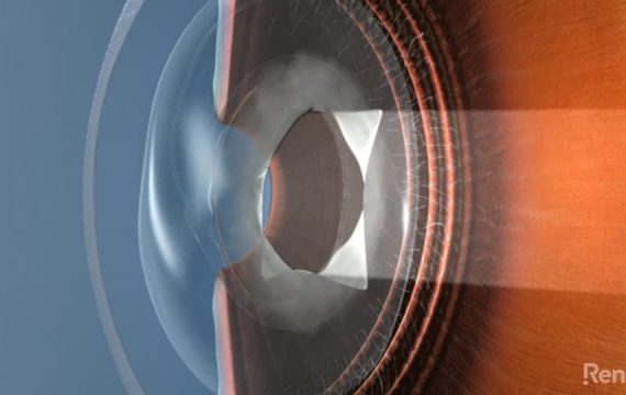
Freedom Eye Laser perform ASLA/ PRK when someone is not suitable for LASIK. LASIK is the preferable procedure due to next day visual recovery and significantly reduced discomfort. However, if LASIK is not appropriate for the individual (given their cornea is too thin or has an abnormal shape) PRK is the treatment of choice. At Freedom Eye Laser we perform Contoura Vision ASLA/ PRK using the Alcon Refractive Suite. As the advanced treatment program for reshaping the cornea is the same as Contoura Vision LASIK, the end result will provide the same excellent visual quality. The procedure involves the front layer of cells of the cornea being smoothed away, with the laser treatment applied directly to the front surface of the eye (as opposed to under a flap as with LASIK). The procedure takes just 15 minutes for both eyes, then a contact lens is positioned and remains in place for 7 days to allow the surface to heal. At day 4 the vision is generally adequate for driving and using a computer. Your vision progressively improves as the cornea remodels its surface layer, with extremely crisp vision taking up to a month.
Blepharoplasty
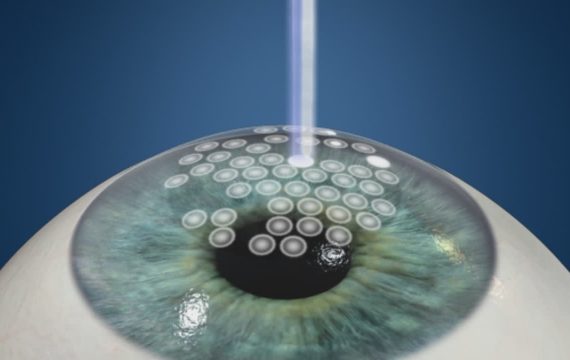
Drooping of the eyelids not only makes you look tired and old but can be very fatiguing and impede your vision. Reducing the excess skin of the upper lid creates a feeling of lightness, opens your vision and helps regain a youthful appearance. Prior to the procedure, the upper lid crease is marked and the redundant upper lid skin is mapped out. This ensures excellent symmetry and contour. The procedure is performed under sedation and takes 45 minutes. There is minimal discomfort, although mild soft tissue swelling and occasional bruising are part of the recovery process. Super-fine sutures are removed painlessly 6 days after surgery. The goal is to ensure a natural look that refreshes both your appearance and vision.
Cataract Surgery - 65 years +
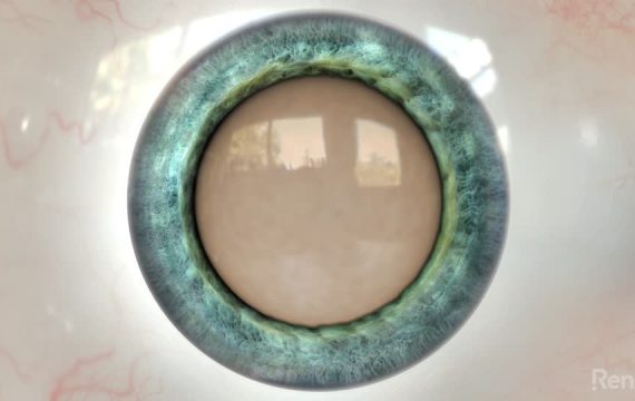
Most people beyond the age of 60 years have some degree of cataract, resulting in a reduction of visual quality. As the natural lens ages, it goes from crystal clear and flexible, to hard and cloudy. The procedure for removing cataracts is to replace the clouding natural lens with a clear artificial implant. Advanced cataract surgery at Freedom Eye Laser uses premium multifocal intraocular lenses to achieve an impressive range of high-quality vision: up close for reading, in between when using computers and sharp in the distance. The spectacular visual results are unattainable by any other means. You will recover clear vision and be free from glasses. Colours leap back to vivid, depth perception is retained and the improvement lasts for life. Find out more...
Collagen Cross-Linking
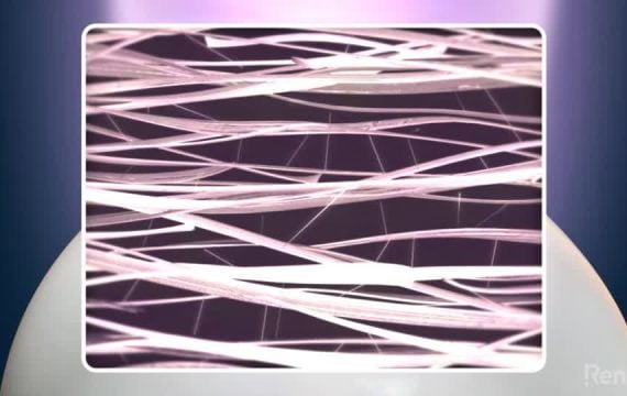
Keratoconus causes the cornea to progressively stretch and bulge, resulting in blurred vision. The only way to halt progression is to perform collagen cross-linking. The procedure takes place in rooms and takes 1 hour. The surface layer of the cornea is disrupted, allowing the riboflavin (vitamin B2) that is dropped onto the eye to absorb into the substance of the cornea. An ultra-violet light of known energy level is then entered on the cornea for 30 minutes. A contact lens is positioned and removed a few days later. The interaction between the UV and the riboflavin allows the collagen fibres of the cornea to link to each other. It stops the progressive steepening of the cornea preventing further visual loss and in some cases improving vision. Advanced topographic linked PRK Laser Eye Surgery can be used in conjunction to further improve vision by normalising the surface curvature.
Corneal ring segments
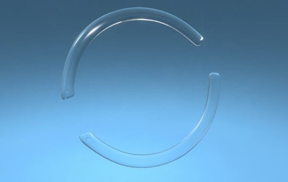
Corneal ring segments are used in treating keratoconus to improve the corneal irregularity and improve vision. The corneal ring segment evens out the shape of the cornea improving both corrected and uncorrected vision. The corneal ring segments are 1mm thick, clear curved crescents of plastic. The 10-minute procedure uses a Femtosecond laser to create a 1mm circular channel within the cornea. The corneal ring segment is then introduced into this channel through a 1 mm incision. Recovery is next day, with vision continuing to improve over several months.
Corneal Transplant - DALK
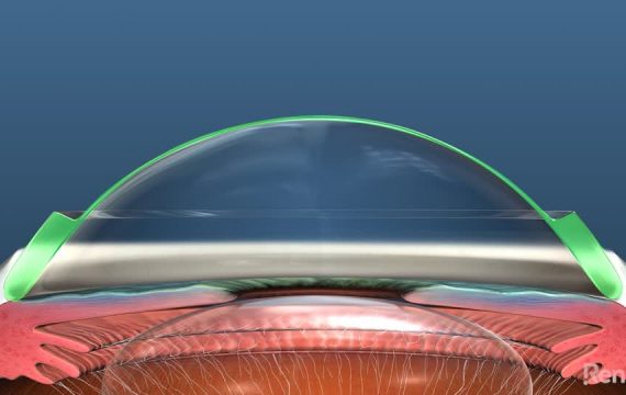
In advanced Keratoconus the abnormal cornea is replaced to provide sharp visual clarity. A DALK is an advanced form of corneal transplantation for keratoconus where the abnormal corneal tissue is removed. The patient’s own endothelium (the cornea’s inner lining) is preserved. By preserving the patient's own endothelium, corneal transplant rejection is eliminated. Using an air bubble (Big Bubble technique) to separate and push back the inner healthy corneal endothelium, only the abnormal corneal tissue is removed. A donor graft is sutured into position. Clarity of vision improves over a month and the sutures are removed at 9 months. Once the DALK transplant settles, the new healthy cornea can be fine-tuned to achieve excellent vision free from glasses or contact lenses.
Corneal Transplant - DMEK
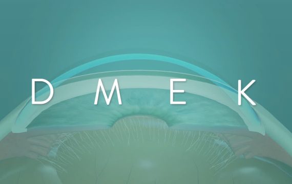
In Fuchs Endothelial Dystrophy vision drops due to hydration of the cornea. The inner lining of the cornea, the endothelium, degenerates and cannot adequately pump fluid out of the cornea back inside the eye. The cornea becomes waterlogged and loses transparency. A DMEK is an advanced form of corneal transplantation where only the diseased tissue is replaced and the remainder of the healthy cornea is maintained. The abnormal endothelium is removed and just the thin membrane of donated corneal tissue with new endothelium cells attached is harvested from the donor cornea, introduced into the host eye and positioned with an air bubble that allows it to adhere to the inner cornea. The cornea can then dehydrate and restore visual clarity. The procedure has a far more rapid recovery and reduced rejection risk compared to traditional corneal transplantation for Fuchs Endothelial Dystrophy.
Focal Macular Laser
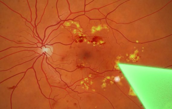
Focal macular laser is used as an adjunct to Intravitreal Injections to assist in resolving a fluid swollen retina in conditions such as Diabetic Retinopathy and a Branch Retinal Vein Occlusions. The procedure is performed in the examinations chair, is discomfort free and takes 5 minutes. After instilling a local unaesthetic drop, a contact lens is positioned. The laser energy is gentle and targeted to the area of swelling. There is no recovery time and the effect of the laser is progressive over weeks.
Intense Pulsed Light (IPL)
83% of Dry Eye is due to Meibomian gland dysfunction (MGD). MGD is a chronic, debilitating disease, which affects millions of people worldwide. It is very irritating and difficult to control through conservative measures. Intense Pulsed Light (IPL) is a recent innovation for the treatment of MGD. Clinical trials demonstrated that 93% of patients improved their tear film quality after 4 treatments of IPL. Many patients improve with IPL where they did not with other therapies. IPL shrinks small blood vessels on the lid margin, reducing inflammation associated with Meibomian Gland Dysfunction. Significant improvements in the tear film stability and dry eye allow the eye to be more comfortable with reduced irritation. The procedure is rapid, safe and comfortable. It is performed as a 10 minute, in rooms treatment with no recovery. Research and clinical experience suggests four treatments, four weeks apart are required for optimal results.
Intraocular Collamer Lens (ICL)
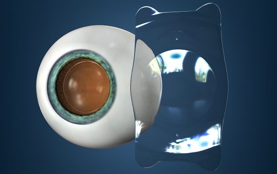
For those with a very large glasses prescription or an abnormal cornea that are unsuitable for laser eye surgery, freedom from glasses and contact lenses can still be achieved with an Intraocular Collamer Lens (ICL). An ICL is a lens that is implanted into the eye that is custom made for the individual. It can correct almost any glasses prescription, regardless of the magnitude or amount of astigmatism. ICL's allow patients under the age of 45 years with corrections that were previously regarded as untreatable, to see perfectly at all distances without glasses. The procedure takes 15 minutes, is pain-free and provides next day recovery.
Intravitreal Injections
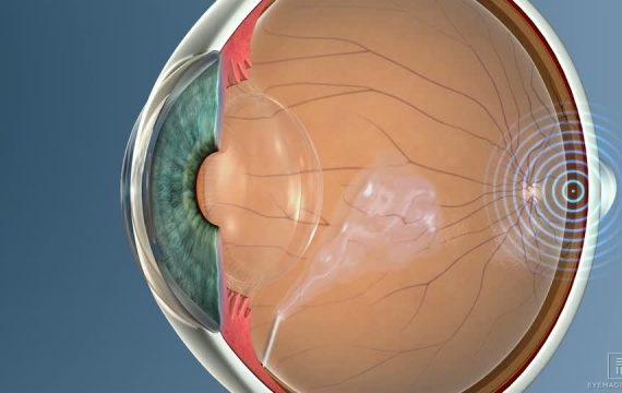
In both ‘wet’ Age-Related Macular Degeneration (ARMD) and Diabetic Retinopathy, fluid swells in the macular - the area of the retina that provides fine central vision. Macular swelling reduces vision and can result in permanent damage if left untreated. Latest generation medications allow the fluid and abnormal blood vessels to regress, leading to stable, improved vision. Intravitreal Injections are a 5 minute, in room procedure performed under local anaesthetic. In the setting of ARMD, a sequence of injections are often 1 to 2 months apart. Although ongoing treatment is often required, conditions such as ARMD and diabetic retinopathy that previously would have resulted in devastating visual loss are now able to be stabilised and often improved.
iStent
iStents are implants that reduce the intraocular pressure for people with Glaucoma. An iStent is a 1mm titanium tube, the smallest implant used in human surgery. By assisting the drainage of fluid inside the eye, it lowers the pressure and protects against the progression of Glaucoma. The procedure is safe and only takes 5 minutes and is generally performed in conjunction with cataract surgery.
Laser Retinopexy
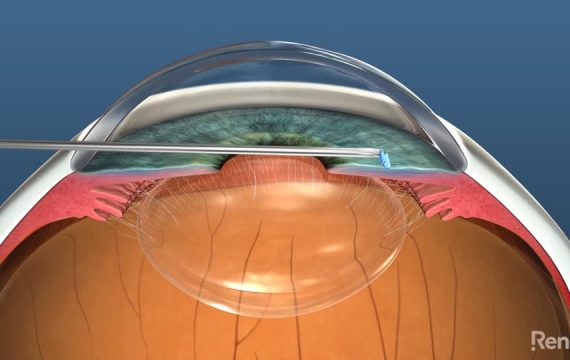
In the uncommon scenario of a Posterior Vitreous Detachment (PVD) resulting in a retinal tear, prompt treatment with Laser Photocoagulation Retinopexy will stop the serious complication of a retinal detachment. After a local anaesthetic drop is instilled, a contact lens is placed on the eye and the laser is performed in an examination chair. Three rows of laser spots surround the tear to stop the influx of sub-retinal fluid, thus preventing retinal detachment. We then reexamine to ensure efficacy, although the treatment is almost always definitive.
LASIK - 20 to 45 years
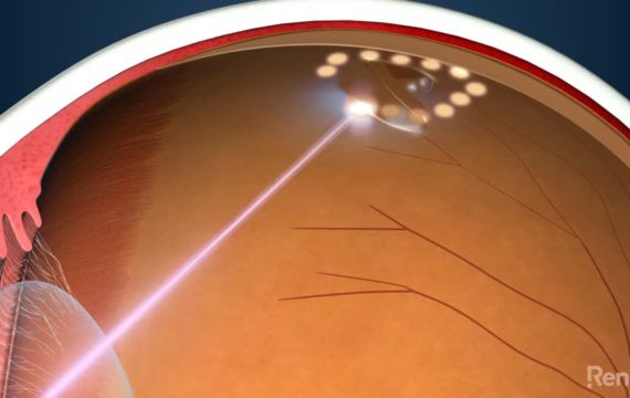
Lasik is an advanced laser vision correction procedure that changes the shape of the cornea to allow freedom from glasses and contact lenses. It is the safest and most accurate procedure performed and can provide vision better than that achievable with glasses or contact lenses. The procedure takes only minutes and provides next day recovery. Contoura Vision LASIK is the pinnacle of laser eye surgery for technology, safety, and exceptional results. We offer the most technologically advanced form of LASIK available to date. Discover how Freedom Eye Laser offer tailored solutions to achieve your best vision possible - Find out more...
Pinguecula Excision
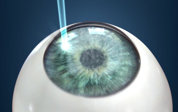
Pingueculum surgery performed at Freedom Eye Laser is considered the gold standard technique. The procedure is performed under an anaesthetic block and takes 30 minutes. The Pinguecula is carefully dissected off the sclera (the white wall of the eye). A free graft of the thin conjunctival membrane from the surface of the white of the eye, hidden underneath the upper lid, is size matched and rotated into the space created on the scleral surface and sutured in with the smallest, dissolvable, sutures used in human surgery. The eye is patched overnight. The use of the anaesthetic block eliminates a large proportion of the discomfort typically associated with Pinguecula surgery. Vision recovery is the next day. Placing a patch graft almost eliminates the risk of recurrence and, once the eye has settled, provides an optimal cosmetic outcome.
PRP - Pan Retinal Photocoagulation
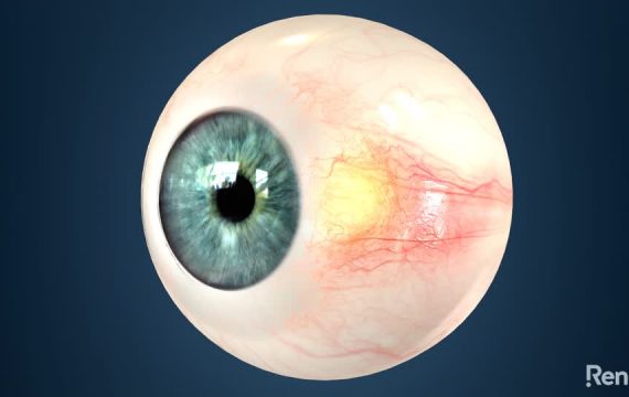
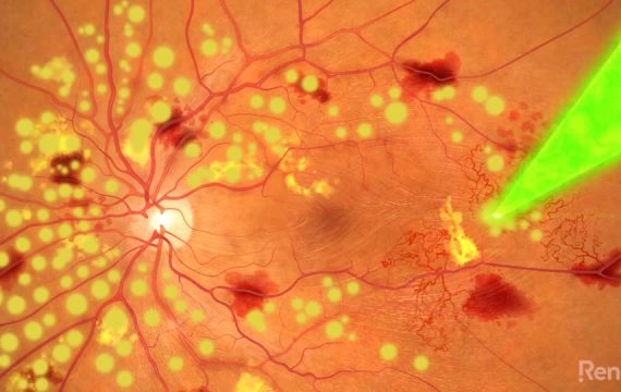
When Diabetic Retinopathy progresses to the advanced state of new blood vessel growth, prompt treatment with PRP can allow serious complications such as vitreous haemorrhage and retinal detachment to be avoided. After local anaesthetic is instilled a contact lens is placed on the eye and the laser is performed in an examination chair. As a large number of laser spots are required to achieve the desired regression of the abnormal blood vessels, the procedure is either performed in multiple instalments determined by the patient’s tolerance to the discomfort or under an anaesthetic local block to numb the eye. There is no recovery time and the effect of the laser is progressive over weeks.
Pterygium Excision
Pterygium surgery performed at Freedom Eye Laser is considered the gold standard. It provides the best cosmetic result and we have never experienced a recurrence over thousands of cases. The procedure takes 30 minutes under an anaesthetic block. The Pterygium is carefully dissected off the cornea and sclera (the white wall of the eye). A free graft of the thin conjunctival membrane from the surface of the white of the eye, hidden underneath the upper lid, is size matched and rotated into the space created on the scleral surface and sutured in with the smallest, dissolvable, sutures used in human surgery. The eye is patched overnight. The use of the anaesthetic block eliminates a large proportion of the discomfort typically associated with pterygium surgery and recovery of vision is next day. By placing a patch graft, the risk of recurrence is almost eliminated and once the eye has settled, the cosmetic appearance is white and normal.
Refractive Lens Exchange - 45 years +
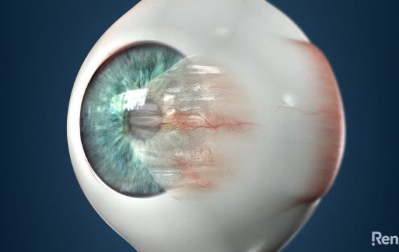
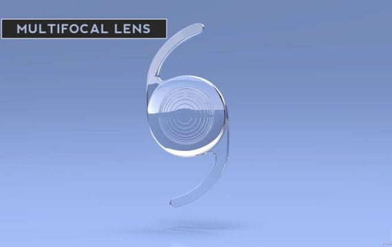
Between the ages of 40 and 50 years, we all start to struggle with near vision due a condition called Presbyopia. Presbyopia literally means “aging eye” – the lens thickens and becomes more rigid, losing the ability to focus up close, particularly in low light. Independence from glasses can be achieved through a refractive lens exchange (RLE) with premium multifocal lenses. Premium multifocal lenses can provide crisp vision at all distances – far, near and at intermediate range. Depth perception is maintained, with both eyes working together as nature intended. The RLE procedure is very similar to cataract surgery. While the lens has aged and is rigid, it has not yet become cloudy. By exchanging the component deteriorating with time, you will never develop cataracts and the exceptional visual results will last for life. Find out more...
Selective Laser Trabeculoplasty
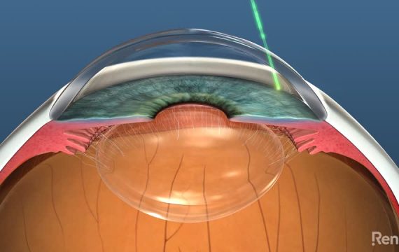
Selective Laser Trabeculoplasty (SLT) is an alternative to drops for first-line glaucoma management. It uses a gentle laser to stimulate the outflow area of the eye to lower the intraocular pressure. The laser is non-tissue destructive, the procedure is painless and takes 5 minutes in the office. It is also generally effective and lasts for around 5 years at which time it can be repeated. SLT can be used as a stand-alone therapy or in conjunction with other glaucoma therapeutic interventions such as drops.
Trabeculectomy
Trabeculectomy is a surgical procedure for glaucoma that is reserved for when control with drops, Selective Laser Trabeculoplasty (SLT) and iStent implantation has been inadequate to halt progression.
During a trabeculectomy, a slow release valve is created utilising a Mini-Express glaucoma shunt to allow a controlled release of fluid from the eye to under the outermost layer of the white of the eye, still isolated from the external environment. Careful follow up is critical. Commonly, the pressure control is effective enough to allow the cessation of medication.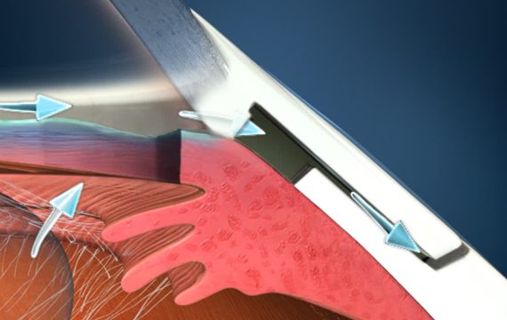
Vitreolysis
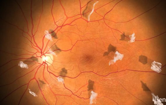
Vitreolysis is a highly effective, outpatient-based laser procedure that aims to remove or reduce floater(s) to a size that no longer impedes vision. The vitreolysis procedure is safe, pain-free and takes around 20 minutes. After local anaesthetic is instilled, a contact lens is placed on the eye and the laser is performed in an examination chair. The laser has the capacity to focus only on the floater, not disturbing anything in front or behind it. The floater is fragmented and vaporised. Generally, a significant improvement is noticed after a single treatment. A second treatment can be performed after 4 weeks if required. The only alternative to resolve symptomatic floaters is to undergo a Vitrectomy - an in-theatre operation with potential complications that are not applicable to Laser Vitreolysis.
YAG Laser Capsulotomy
In advanced cataract surgery, premium intraocular lens implants are used to achieve glasses-free, youthful vision. The lens is implanted within the capsule that used to encompass the natural lens. As a healing reaction, it is common that the capsule will start to thicken over time affecting clarity of vision. A YAG Laser Capsulotomy quickly and permanently resolves this. The procedure is safe, pain-free and takes around 5 minutes in rooms. After local anaesthetic drop is instilled, a contact lens is placed on the eye and the laser is performed in an examination chair. The laser has the capacity to focus only on the thickened capsule and clears the visual axis, returning the vision to its optimal state immediately. The YAG Laser Capsulotomy never needs to be repeated and is free of risk and recovery.
